/ THE SHORT ANSWER
- 01Identify the specimen and anatomical coverage before comparing maps.
- 02Separate the dataset release from the later journal publication.
- 03Treat a wiring diagram as the starting point for functional experiments.
/ dotSuper point of view
The advance is a more complete experimental reference, not evidence that biological intelligence has been copied into software.
A map that connects the brain to the nerve cord
HHMI Janelia led the project with Cambridge researchers, Google Research and collaborators.
The associated study appeared in Cell.
The anatomical coverage matters as much as the headline count.
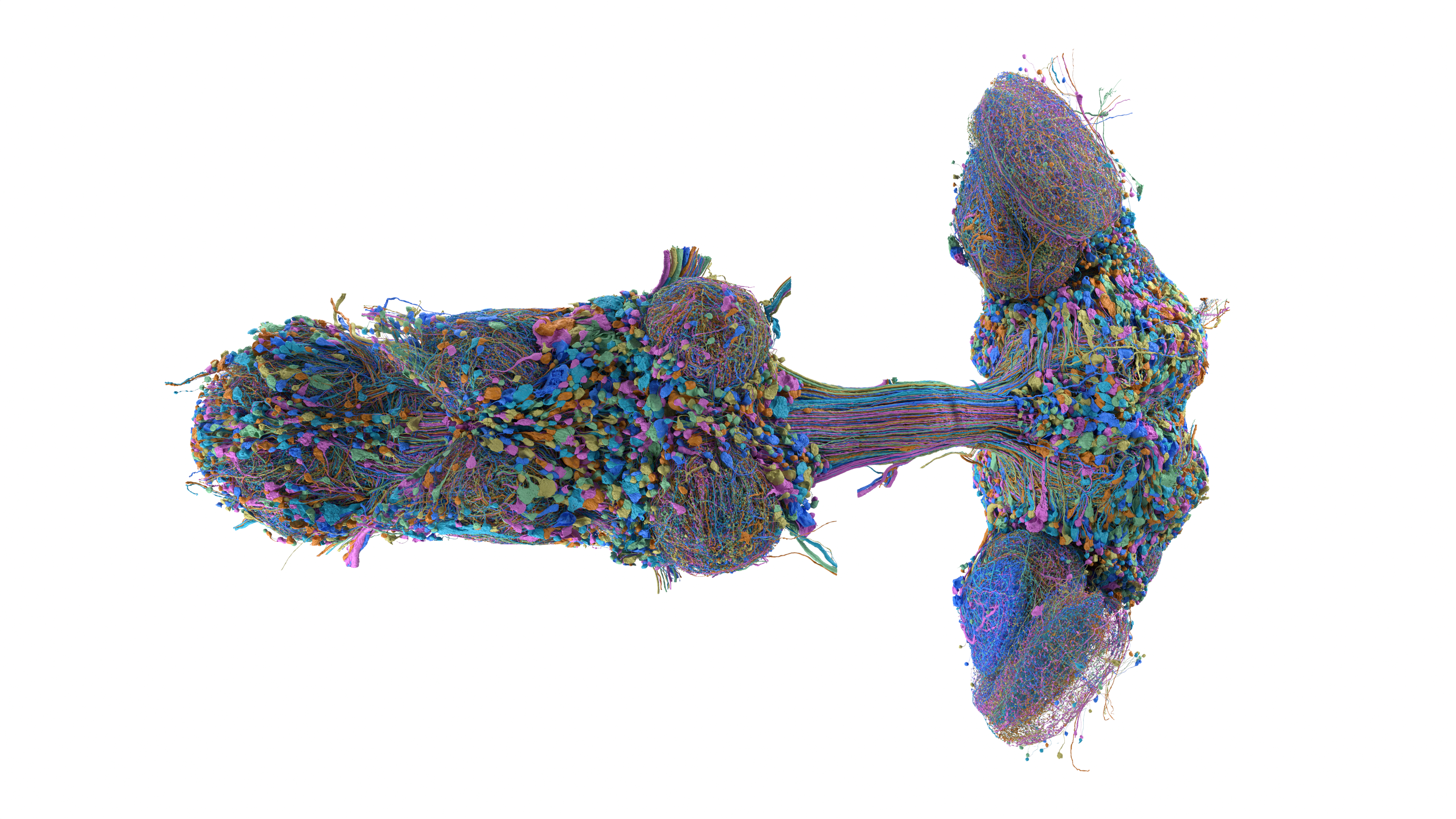
MaleCNS includes central brain, optic lobes and the ventral nerve cord.
The nerve cord is involved in controlling the body; it is not interchangeable with the brain.
A useful first question is therefore not simply 'How big is the map?'
Ask whether a circuit can be followed across the anatomical boundary relevant to the experiment.
Why the dates in different headlines disagree
The journal publication and a new wave of public coverage followed in September.
Earlier access to a dataset and later publication are different milestones.
A video posted this week might show a resource that researchers have used for months.
That does not make the science less important.
It changes what is genuinely new in the story.
When sharing this research, record three things separately: dataset version, publication date and demonstration date.
This small habit prevents an old simulation from being presented as a new biological discovery.
| Milestone | What it establishes |
|---|---|
| Dataset release | The version of the map available for analysis |
| Journal publication | The research record and accompanying methods |
| Later demonstration | A particular use or visualization of the resource |
How this differs from the 2024 female-brain map
That paper describes 139,255 neurons and 54.5 million synapses between them.
Do not turn those numbers into a male-versus-female intelligence comparison.
MaleCNS and the 2024 FlyWire resource cover different anatomical extents.
Later female CNS work also exists, so 'female map' is not one timeless dataset.
For any comparison, match the tissue, filtering rules, reconstruction versions and cell categories.
Otherwise a larger total may reflect what was included rather than a biological explanation.
How to read the research image
It shows representative neuron types rather than a natural-color photograph of a living brain.
Colors help distinguish reconstructed structures.
They do not demonstrate that a neuron is firing, reveal a memory or indicate a measured emotional state.
Follow the image credit to the research gallery for its original description.
A striking visualization becomes more useful when the reader knows what was selected and what the colors mean.
What this makes possible, and what it does not
Those are concrete research uses, even before a complete account of behavior is available.
Our interpretation is that this milestone should raise the standard for explanations, not lower it.
A claim about behavior needs more than a colorful network or a neuron count.
For a practical reading exercise, choose one claimed behavior and write down the proposed input, intermediate circuit and output.
Then ask what recording or intervention would distinguish that explanation from an alternative.
- Read the project overview before watching viral demonstrations.
- Keep the map, model and measured behavior as separate evidence layers.
- Use the companion guides to explore simulations, applications and limitations.
What this page cannot conclude
- 01Numbers refer to specific resources and versions, not universal neuron totals for every fly.
- 02The rendering is structural. Neither it nor the map establishes consciousness, remembered experience or a complete behavioral simulation.
- 03Prepared with AI assistance from the named research sources. dotSuper did not conduct these experiments, independently reproduce the studies or obtain an endorsement from the research institutions.
Sources
- 01A connectomics milestone: mapping the complete male fruit fly brainGoogle Research · accessed Sep 15, 2026
- 02Sexual dimorphism in the complete Drosophila male central nervous system connectomeBerg et al., Cell (2026), PubMed record · accessed Sep 15, 2026
- 03MaleCNS release notesMaleCNS project · accessed Sep 15, 2026
- 04Neuronal wiring diagram of an adult brainDorkenwald et al., Nature (2024) · accessed Sep 15, 2026
- 05Male CNS Connectome: project overviewHHMI Janelia / FlyEM · accessed Sep 15, 2026
- 06MaleCNS research media galleryFlyEM / HHMI Janelia and collaborators · accessed Sep 15, 2026
Our editorial standard · Found an error? Send a correction with its source.
/ CITE OR SHARE THIS GUIDE
Make the evidence easy to verify.
When you reference this guide, link to its canonical URL. That gives readers one stable place for the evidence, limitations and future updates.
dotSuper Research Desk. (September 15, 2026). The New Fruit Fly Brain Map, Explained. dotSuper. https://dotsuper.net/feeds/applied-systems/fruit-fly-brain-map-2026-explained
What Is a Connectome, and What Is Missing?
Continue with a different question in our source-linked fly-brain series.
Read the next explainer